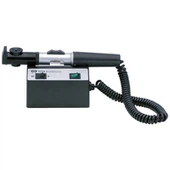
直接 検眼鏡 缶 直接 検査 眼底 なし 拡張 瞳孔 検査 is 運ばれた 出た in a 暗い 部屋. 検査者's 目 必見 be close to 患者's 目, check the 患者's 右 目 あり 右 目, ホールド the 検眼鏡 in the right hand of the 患者, 座る または 立つ on the right side of the patient, and the left eye is vice versa。 使用 +10D レンズ to check かどうか the 患者's 屈折 間質 is 透明, 後 チェック the 屈折 間質, you can begin to 検査 すべて 部分 of the 眼底, rotate the disc of the lens to correct the doctor and the patient's refractive errors, if the doctor is emmethean or has been equipped with corrective glasses, the diopter used to see the fundus indicates the refractive condition の 検査 目。
一般的に, the 影響を受けた 目 is looked まっすぐ 前方, the 光学 乳頭 is 検査済み, and then the superior 側頭, infratemporal, superior 鼻, and 劣った 鼻 象限 are 検査済み に沿って 大中 血管. 最後に, the 影響を受けた 目 is 固定 on the 側頭 側, and the 黄斑 is 検査済み. The size of the fundus 病変 was 発現 by the 直径 of the optic nipple, and the degree of 曲率 of the 病変 was measured by the 視度 of the lens. 3D was 同等 to 1mm. Some 検眼鏡 are 装備 with green filters for better 観察 of 光学 神経 線維 および 黄斑。
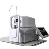
When the indirect ophthalmoscope is used, the pupil must be fully dilated and examined in the dark room. The doctor should switch on the power supply, adjust the distance and the position of the mirror, observe with weak light first, see the opacity of the cornea, crystal and vitreous body, and then direct the light into the pupil of the examined eye, and let the examined eye focus on the light source. Generally, the +20D objective lens is placed at 5cm in front of the examined eye. The convex surface of the objective lens faces the examiner, the examiner holds the objective lens with his left hand, and fixes it on the orbital margin of the patient. The examined eye, the objective lens, and the head of the examiner are fixed. When the optical papilla and macula are seen, the objective lens is moved to the direction of the examiner, and the stereoscopic inversion of the optical papilla and macula can be clearly seen at 5cm in front of the subject.
When examining the rest of the fundus, the subject should be able to move his eyes to cooperate with the examination, and the examiner should move around the subject's head, and the hand-held objective and the examiner's head should move accordingly. The images are opposite up and down, opposite left and right. To examine the periphery of the fundus, such as the 6 o 'clock position, the examiner is located on the top of the client's head, so that the affected eye looks down at the 6 o 'clock position. The examination of the far peripheral part of the fundus must be combined with scleral compression method. The metal scleral compression device is worn on the middle or index finger of the right hand of the inspector, and the head of the compression device is placed outside the corresponding eyelid of the examined eye. If necessary, the examination can be carried out from the conjunctival sac after epimanaesthesia. During the operation, the line of sight of the examiner should be kept in a straight line with the illumination of the indirect ophthalmoscope, the focus of the objective lens, the inspected eye position, and the head of the oppressor. During the examination, attention should be paid to asking the patient to close the eyelid at any time to wet the cornea. When there is a suspected intraocular space occupying lesion, pressure examination should be avoided.
タイプ および アプリケーション メソッド of 検眼鏡
Aug 21, 2023
伝言を残す
あなたはおそらくそれも好きでしょう